

Global win for Australian retinal image
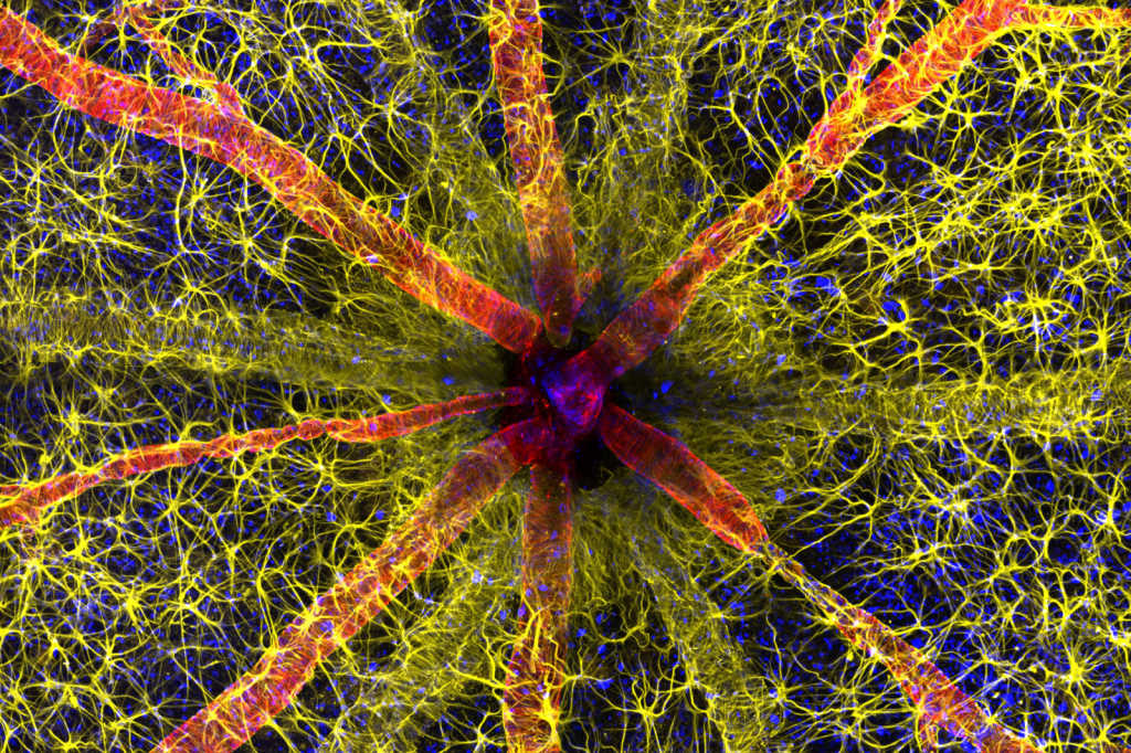
Two Lions Eye Institute researchers have won this year’s Nikon Small World Photomicrography Competition with a detailed image of a rodent’s optic nerve head.
Student Hassanain Qambari and research assistant Jayden Dickson’s photograph highlighted the intricacies of retinal vasculature, with astrocytes shown in yellow and contractile protein in red. Qambari, who is investigating ways to detect diabetic retinopathy, said he thought the competition would be a great way to show the retina’s intricacies and why the study of retinal diseases poses such a challenge. The pair were awarded US$3,000 (NZ$5,000).
The researchers stained the vasculature via cannulation and perfusion of the ophthalmic artery (100-120µm diameter), a technique pioneered by the Lions Eye Institute Physiology and Pharmacology research team more than 20 years ago. The retina was then dissected, separating it from the sclera and choroid. Since intravascular perfusion cannot reach astrocytes, that part of the retina was immersion stained. A Nikon Confocal microscope under a 20x objective lens then captured the image, with lasers of different wavelengths used to excite molecules bound to the cellular markers of interest.
Related Articles
-
Fixing fixation for more accurate perimetryDecember 22, 2023
-
2023 – a year of firsts and returning favouritesDecember 21, 2023

-
Promising Stargardt therapy revealedDecember 20, 2023
-
Poster highlights: a snapshot of studies at RANZCO 2023December 20, 2023
-
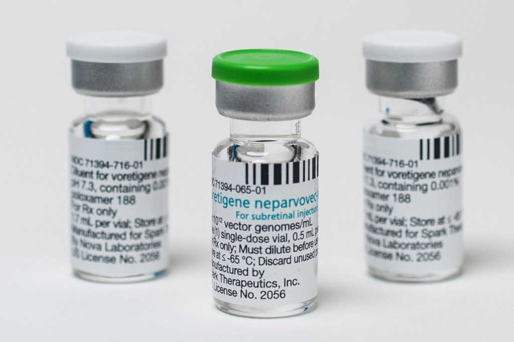
Luxturna funding application stallsDecember 19, 2023
-
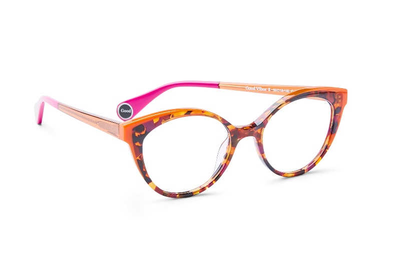
Exuding good vibesDecember 19, 2023
-
Maintaining eye standards in ANZDecember 18, 2023

-
Success for novel DED dropDecember 17, 2023